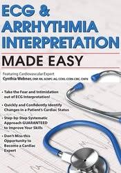
ECG & ARRHYTHMIA INTERPRETATION MADE EASY – CYNTHIA L. WEBNER
- Take the Fear and Intimidation out of ECG Interpretation!
- Quickly and Confidently Identify Changes in a Patient’s Cardiac Status
- Step-by-Step Systematic Approach GUARANTEED to Improve Your Skills
- Don’t Miss this Opportunity to Become a Cardiac Expert
The ECG is the most commonly-used procedure for diagnosing a wide variety of cardiac conditions, yet even the most seasoned nurses struggle to master interpretation. Attend this seminar and finally learn to recognize borderline and abnormal tracings. In this information-packed program expert, Cynthia L. Webner, will teach you easy-to-understand techniques to identify acute coronary syndromes, bundle branch blocks and challenging rhythms you would have previously missed. Through hands-on practice, learn how to look at and analyze:
- QRS axis to help determine diagnosis
- Contrast left anterior and posterior hemiblocks
- Recognize the location of acute coronary syndromes
- Differentiate LBBB versus RBBB and their significance
- Find chamber enlargements
- And most importantly, respond to the patient emergencies associated with each of these changes!
- Choose correct electrode placement required for accurate 12-lead ECG acquisition.
- Assess normal and abnormal patterns on each lead of the 12-lead ECG.
- Determine cardiac axis using lead I and aVF.
- Analyze the 12-lead ECG features seen in atrial and ventricular hypertrophy.
- Specify the features of right bundle branch block from the features of left bundle branch block.
- Evaluate patterns of infarct, injury, and ischemia on the 12-lead.
- Utilize morphology in Lead V1 and V6 to differentiate ventricular tachycardia from SVT with aberrant conduction.
- Categorize high risk features for Torsades de Pointes and other cardiac arrhythmias.
Time Saving Tips & Strategies
- Systematic Approach
- Correlating Cardiac Conduction with Waveforms
- Layout of the 12-Lead, 15-Lead & Right-sided ECG
- Positive & Negative Lead Placement
- Cardiac Conduction System Clues
Determining Cardiac Axis
- Quick Approach for Axis by Quadrant
- Axis Practice Utilizing “Thumbs Technique”
- Causes for Axis Deviation
- Axis in Disease Diagnosis
Conduction Abnormalities
- Right & Left BBB Morphology
- Left Anterior Hemi-Block Criteria
- Chamber Enlargement
- Atrial Hypertrophy
- Right- & Left-Ventricular Hypertrophy
Myocardial Injury and Ischemia
- Patterns of Injury & Ischemia
- ST Segment & T Wave Changes
- Reciprocal Changes
- Pathological Q waves
- Specific Types of Myocardial Infarction
- Subtle Clues
Complex Arrhythmia Interpretation
- Mechanisms of Tachyarrhythmias
- Evaluating Wide Complex Tachycardias
- VT versus SVT with BBB Aberrancy
High Risk Features
- QT abnormalities
- The Brugada Syndrome
- Wolff Parkinson White Syndrome
Putting It All Together
- Emergency Interventions
- Acute vs. Chronic Treatment Recommendations
- Documentation of Findings
Tag: ECG & Arrhythmia Interpretation Made Easy – Cynthia L. Webner Review. ECG & Arrhythmia Interpretation Made Easy – Cynthia L. Webner download. ECG & Arrhythmia Interpretation Made Easy – Cynthia L. Webner discount.

